


To the Editors:
Bilateral pulmonary nodules are a relatively common finding in thoracic radiology and hence are an important problem that chest physicians often face. Aetiology can be diverse, ranging from neoplastic conditions to infectious lesions. An accurate and prompt diagnosis is needed in order to target the most appropriate and least harmful treatment. We report the case of a 49-yr-old, Caucasian, otherwise healthy male patient with bilateral lung nodules and an unusual diagnosis.
The patient had an unremarkable history and was referred due to asymptomatic bilateral lung nodules. He had stopped smoking 6 months earlier and had a normal chest radiograph a year before referral, taken for follow-up of benign colon polyps. At the time of presentation he was not on any medication.
The patient was an engineer working in a Belgian high-tech facility. His job mainly consisted of office work with occasional visits to production sites where coating of medical material was performed. When visiting these sites, he did not systematically wear a protective mask. The problems occurred after a business trip to the USA where he met with three colleagues. He first visited his general practitioner with a flu-like syndrome. Other symptoms included a productive cough, night sweats and loss of weight (5 kg in 2 weeks). He was treated symptomatically with paracetamol. Two of his colleagues developed the same complaints. One never visited a doctor but the other did and was diagnosed with sarcoidosis based on the finding of non-necrotising granulomatous lesions in transbronchial biopsies. He was put on steroids and his condition improved. Oddly, the third colleague who did not visit the doctor had an existing diagnosis of sarcoidosis but remained asymptomatic.
Due to persistent cough and low-grade fever, our patient was subsequently referred to a pneumologist. Biochemistry revealed an inflammatory syndrome (C-reactive protein 4.6 mg·dL−1, sedimentation rate 28 mm·h−1) and normal tumour markers, angiotensin-converting enzyme (ACE) levels and anti-nuclear factor (ANF). Mycoplasma pneumoniae serology was compatible with a recent infection. Computed tomography (CT) of the chest showed bilateral lung nodules, some with cavitation, in a subpleural and more central distribution. Extensive mediastinal lymphadenopathy was also present. Bronchoscopy revealed a normal bronchial tree. Bronchoalveolar lavage was neutrophilic (60%) with no bacterial pathogens and a normal cytology. Pulmonary function tests were normal (vital capacity 4.9 L, forced expiratory volume in 1 s (FEV1) 4.4 L, FEV1/forced vital capacity (FVC) 88%, total lung capacity 9.2 L, diffusion capacity 99%). Atypical pneumonia was diagnosed and the patient was treated with doxycycline 200 mg daily for 10 days.
2 months after initial presentation the symptoms disappeared and biochemistry normalised. The lung nodules persisted but the cavitations and adenopathy regressed. The patient was then referred to our centre for a second opinion. At that time he was completely asymptomatic and the initial weight lost had been regained. Our differential diagnosis included chronic berylliosis, sarcoidosis and, less likely, malignancy and opportunistic infections. A lymphocyte transformation test (LTT) performed for berylliosis was negative. Bronchoscopy with lavage, brushing and biopsies was repeated and showed a normal cell count and differentiation, a CD4/CD8 ratio of 3.8 and no bacterial pathogens on cultures. Bronchial brushing and biopsies were normal. HIV test was negative.
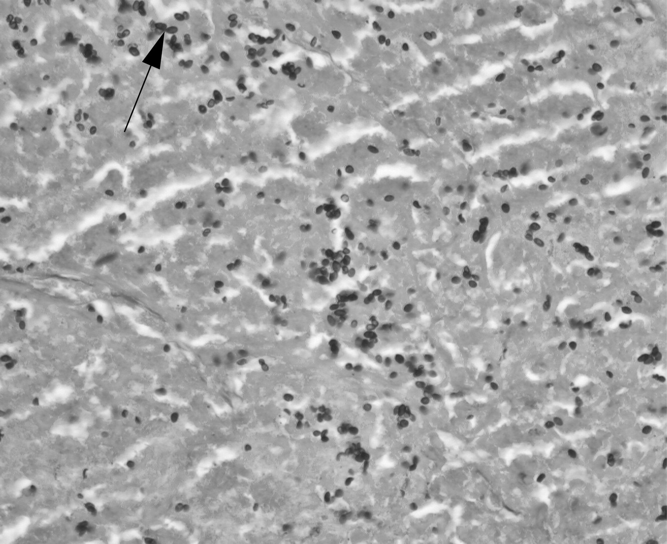
5 months after his business trip the patient remained asymptomatic. No further change of the radiological picture was noted. Exposure to beryllium seemed very unlikely since the LTT was negative. Given the uncertainty of the diagnosis, a lung biopsy was performed. This revealed necrotising granulomas with a positive Grocott methenamine silver (GMS) stain (fig. 1). A serological test for Histoplasma capsulatum antibodies confirmed pulmonary histoplasmosis. Our patient did not receive any specific treatment and remained clinically stable 6 months after initial presentation. Intriguingly, retrospective examination of the histopathological samples showed the presence of the same micro-organism in our patient's colleague who was diagnosed with sarcoidosis and treated with steroids. His clinical improvement seemed unexpected as steroids are known to depress the cellular immunity, which would facilitate the infection with H. capsulatum 1. We did not ask to review the transbronchial biopsies of the third colleague as his diagnosis of sarcoidosis was prior to the business trip and he remained asymptomatic thereafter.

Necrotising granuloma from lung biopsy specimen. Small ovoid yeasts, some with narrow-based budding (arrow), representing Histoplasma capsulatum in a background of necrosis. Grocott methenamine silver stain. Original magnification ×200.
Granulomas are amongst the most common pathological findings in pulmonary medicine; yet diagnosis often remains challenging due to incomplete clinical data and the difficulty of interpreting some histological features. Granulomas are compact aggregates of histiocytes (macrophages). They may also contain necrosis (necrotising granuloma), lymphocytes, plasma cells or multinucleated giant cells 2. Infections are the first cause of granulomatous lung disease, with mycobacteria and fungi as the two most common infectious agents. Sarcoidosis is a primary non-infectious cause, but other causes include chronic berrylium disease, hypersensitivity pneumonitis and, less frequenly, hot tub lung and Wegener's granulomatosis 2.
H. capsulatum is a dimorphic fungus existing as a mould in the soil and as a yeast at body temperature 1, 3. Soil containing bat or bird droppings is the reservoir. The fungus is endemic to certain regions such as North and Central America, but also to Africa, Southern Europe and South-Eastern Asia. Exposure in these locations is extremely frequent but symptomatic infection is less common and depends on the balance between cell-mediated immunity and infectious burden. In an immunocompetent host, a large inoculum is needed to cause the disease, while immunocompromised patients are at risk of developing it with a much smaller inoculum. Contamination occurs with inhalation of the microconidia that reach the alveoli and infect the macrophages before spreading via the reticuloendothelial system 1, 3.
Clinical pictures can be acute or chronic, local or disseminated and possibly complicated. The acute pulmonary forms are a mostly mild pneumonia mimicking an atypical pneumonia and are thus often treated as such, as in our patient. Severe forms also exist and can lead to acute respiratory distress syndrome (ARDS). Chronic cavitary pulmonary histoplasmosis occurs generally in patients with emphysema, resembling a reactivation of tuberculosis. Complications of these pulmonary forms include granulomatous mediastinitis, fibrosing mediastinitis and pericarditis, but these are less common. Disseminated histoplasmosis can be either acute, leading to a sepsis-like illness, or chronic. Immunocompromised patients develop acute illness. Chronic disseminated histoplasmosis is seen in older, mostly male patients with underlying emphysema but normal immunity 1.
Diagnosis is via careful review of patient history and can be confirmed using various tests. The test used depends on the clinical syndrome 1, 2, 4. For chronic histoplasmosis, serology is the test of choice. Complement fixation and immunodiffusion techniques can both be used. Antibody production takes 4–8 weeks, however, and repeating the test might be necessary in case of strong clinical suspicion and initial negative serology. False-positive serology can be seen in lymphoma, tuberculosis, sarcoidosis and other fungal infections. A persistent low-positive titre can also be noted years after exposure and therefore does not always correspond to active infection 1. Culture of respiratory specimen is also indicated in chronic pulmonary forms but pathogen growth may take weeks. In acute and particularly disseminated forms, antigen detection is rapid. Urinary testing has the best sensitivity. PCR techniques are not yet reliable. Histopathology can be divided into three main types 2. The first is an intra-alveolar lymphohistiocytic infiltrate with small granulomas and variable necrosis, mainly observed in acute pulmonary histoplasmosis. The second shows well-formed, necrotising granulomas surrounded by a rim of epithelioid histiocytes and a fibrotic capsule. These lesions, also called “histoplasmomas”, are often resected in the work-up of a solitary pulmonary nodule 2, 5. The third histological subtype is mostly observed in disseminated disease and consists of heavily infected histiocytes within the interstitium 2, 6. H. capsulatum are usually not visible in granulomas on haematoxylin–eosin stains but GMS stains enlighten small, uniform, ovoid organisms, sometimes typically narrow-based 1, 4.
Treatment is only proven to be effective in severe pulmonary and disseminated forms. The agent of choice is amphotericin B for the most severe syndromes. It is recommended to use the liposomal form rather than the deoxycholate because of the lower nephrotoxicity. Itraconazole is used as a maintenance drug and in milder cases. Treatment can last from weeks to months, depending on the severity of the infection 1, 7.
In conclusion, this case highlights the importance of pathology and reminds us of the endemically important differential diagnoses for sarcoidosis. It also proves that, in all countries, the first step when analysing lung granulomas should be to rule out infection.
Footnotes
Statement of Interest
None declared.
- ©2011 ERS


